50+ Years — Excellence in Ophthalmology

The best vision possible. Yours.

South Florida’s leader in custom no-flap LASIK, laser cataract surgery, and corneal cross-linking — performed by the Rand physicians in our own surgical pavilion in Deerfield Beach.

Trusted by South Florida families for over 50 years

Rand Eye Institute — 50+ Years, Excellence in Ophthalmology

Request an appointment

We reply within one business day — or call us directly.

Your information is used only to contact you about your care. Please do not send sensitive medical details through this form.

50+
Years of surgical expertise
136,000+
Surgeries performed
35
Years of Joint Commission accreditation
24,000
Square feet dedicated to eye care

Three floors. Twenty-six exam and treatment rooms. Four surgical suites — every stage of your care in one building.

What we do best

Complete eye care.
A tailored approach.

Every eye is different, which is why we never start with the procedure. We start with your measurements, your health, and how you actually use your vision.

Complete Eye Health

Comprehensive Ophthalmology

Full medical eye care for adults — examinations, diagnosis and ongoing management of the conditions that affect how you see, from routine vision changes to complex disease.

  • Complete diagnostic examination
  • Ongoing management of chronic eye conditions
  • Referral within the practice when a specialist is needed
See if you’re a candidate

Premium Lens Implants

Refractive Cataract Surgery

Cataract surgery is no longer only about removing a cloudy lens. With premium lens implants it becomes an opportunity to address a lifetime of prescription at the same time.

  • LenSx® laser-assisted, computer-guided precision
  • PanOptix® trifocal lens options
  • Distance, near and astigmatism addressed together
  • Outpatient procedure in our own surgical pavilion
See if you’re a candidate

Custom No-Flap LASIK

Laser Vision Correction

Wake up, look at the clock, and simply see. Our custom, no-flap laser vision correction is mapped to the unique optical fingerprint of your eye — not to an average.

  • No-flap technique — no blade, no corneal flap
  • Wavefront mapping of your individual eye
  • Performed in our on-site Rand Surgical Pavilion
See if you’re a candidate

Minimally Invasive

Corneal Collagen Crosslinking

For keratoconus and corneal thinning, crosslinking strengthens the collagen bonds inside your cornea — a minimally invasive treatment intended to halt progression and protect the sight you have.

  • Minimally invasive, performed in-office
  • Strengthens corneal collagen to stop progression
  • The standard of care for progressive keratoconus
See if you’re a candidate

Ocular Surface

Dry Eye & Ocular Surface Disease

Chronic dry eye is an inflammatory disease of the tear film and ocular surface, not simply a shortage of tears — which is why drops alone so often fall short.

  • Tear film and ocular surface testing
  • Treatment aimed at the underlying inflammation
  • Management of chronic and post-surgical dryness
See if you’re a candidate

Retina

Medical Retina & Injections

Medical management of the retina — macular degeneration, diabetic retinopathy, macular oedema and related disease, including in-office intravitreal injection therapy.

  • Macular degeneration, wet and dry
  • Diabetic retinopathy and macular oedema
  • In-office intravitreal injections
  • Retinal imaging and monitoring over time
See if you’re a candidate

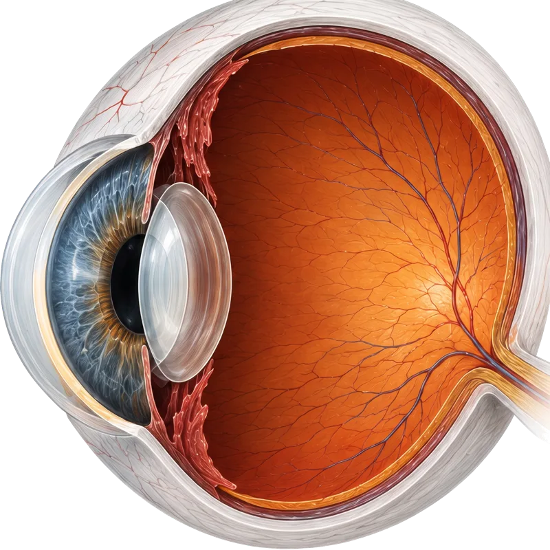
Understand your eye

How sight actually works.

Almost every condition we treat comes down to one of these parts not doing its job. Knowing which one it is makes your own diagnosis far easier to follow.

Cutaway illustration of a human eye
  1. 1CorneaThe clear front window of the eye. It does most of the focusing before light reaches the lens.
  2. 2IrisThe coloured ring. It opens and narrows the pupil to control how much light gets in.
  3. 3PupilThe opening at the centre of the iris. It widens in dim light and shrinks in bright light.
  4. 4LensSits behind the iris and fine-tunes focus. When this lens clouds, that is a cataract.
  5. 5ScleraThe tough white outer wall that protects the eye and holds its shape.
  6. 6ChoroidThe layer of blood vessels between sclera and retina that supplies it with oxygen.
  7. 7RetinaThe light-sensitive lining at the back. It converts light into nerve signals.
  8. 8MaculaThe small central patch of retina responsible for sharp, detailed vision. Macular degeneration affects this.
  9. 9Optic nerveCarries the signal from the retina to the brain. Glaucoma damages these fibres.
  1. 1Light enters the eyeLight passes through the cornea.
  2. 2FocusThe cornea and lens focus it onto the retina.
  3. 3ConversionThe retina turns light into electrical signals.
  4. 4Brain signalThe optic nerve carries them to the brain.

See it for yourself

What you see matters.

Choose a condition, then drag to move between how it can look and a corrected view. For many of our patients this is not a metaphor — it is the week of their procedure.

A sunlit meadow of orange poppies, blue cornflowers and pink cosmos in sharp, vivid focusCataractCorrected

A clouding lens scatters light: colours fade and yellow, contrast drops, and bright light causes glare.

An illustrative simulation, not a diagnosis. How a condition affects your sight — and what can be done about it — is determined by an examination.

Why Rand

Four exam rooms at the start.
One of the largest in the country today.

Dr. William J. Rand opened this practice with one nurse, one optometrist, and two receptionists. What has not changed in fifty years is the belief that the surgeon who recommends a procedure should be the one who performs it — and the one who checks on you afterward.

01

One building. Every step.

Consultation, diagnostics, surgery, and follow-up all happen under one roof, in our own surgical pavilion. You are never handed off to a facility that has never met you.

02

A surgeon, not a schedule.

The physician who evaluates your eyes is the physician who operates on them, and the one who sees you afterward. Continuity is the whole point.

03

Technology chosen, not collected.

We adopt a platform when it measurably improves an outcome — LenSx, PanOptix, premium lens implants — and we train the entire team on it before it touches a patient.

04

Fifty years of judgment.

We have grown from four exam rooms to one of the largest comprehensive eye care facilities in the country, with over 100 professionals on staff.

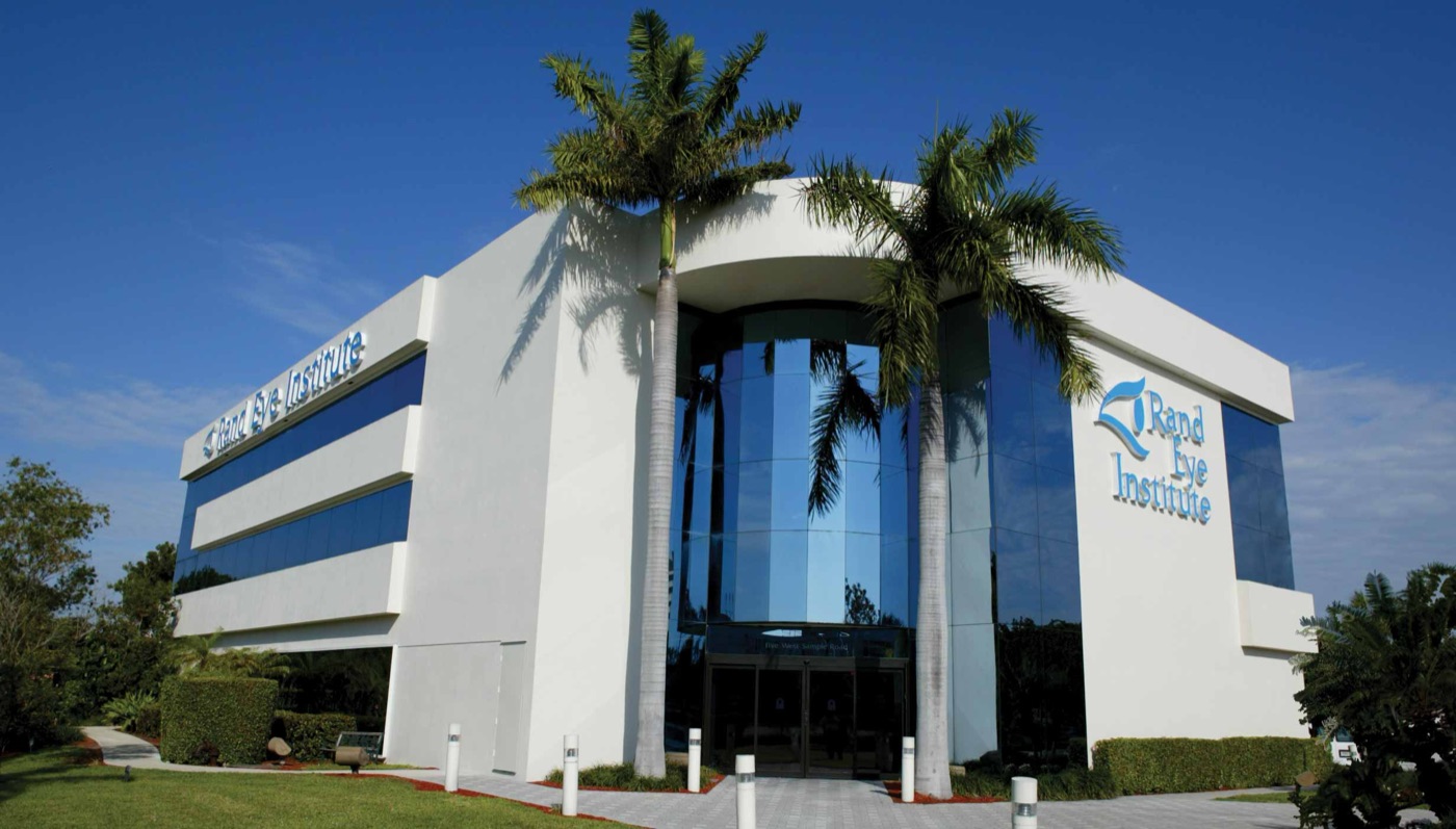
The Rand Eye Institute building on West Sample Road in Deerfield Beach, Florida

The Rand Surgical Pavilion

Three floors, a self-contained operating suite, and every stage of your care in one place — state-licensed and Joint Commission certified.

100+

Eye care professionals under one roof

3

Floors, including our own surgical pavilion

The physicians

Family owned and operated.

The Rand family has led this institute for over 50 years. You will know your surgeon by name — and they will know your eyes by their measurements.

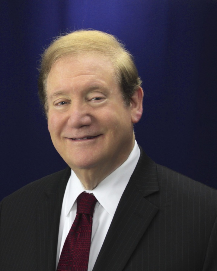
William J. Rand, M.D., Medical Director & Founder at Rand Eye Institute

Clearly focused on the best vision possible.

William J. Rand, M.D.

Founding Physician

Medical Director & Founder

  • Founding Physician, Rand Eye Institute
  • Member, American Academy of Ophthalmology
  • Fellow, American College of Surgeons

Areas of focus

  • Comprehensive Ophthalmology
  • Cataract & Refractive Surgery with Premium Lenses
  • LASIK
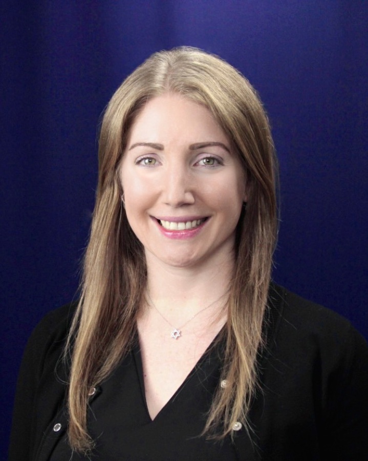
Allison L. Rand, M.D., Cornea · Dry Eye · Refractive Cataract Surgery at Rand Eye Institute

Cornea, cataract, and a lifetime of dry-eye expertise.

Allison L. Rand, M.D.

Fellowship Trained — Cornea

Cornea · Dry Eye · Refractive Cataract Surgery

  • Fellowship: Cornea & External Disease
  • Diplomate, American Board of Ophthalmology
  • Ocular Surface Center Director

Areas of focus

  • Comprehensive Ophthalmology
  • Cataract & Refractive Surgery with Premium Lenses
  • Corneal Transplantation
  • Corneal Collagen Crosslinking
  • LASIK
  • Dry Eye & Ocular Surface Disease
  • Pterygium Surgery
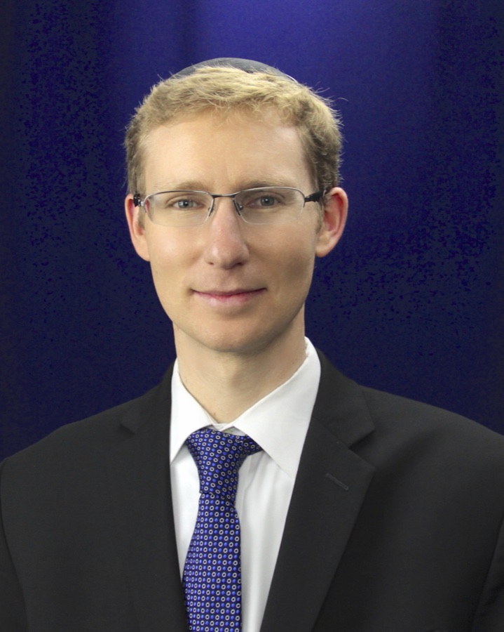
David L. Rand, M.D., Director of Innovation · Refractive & Cataract Surgery at Rand Eye Institute

Bringing LenSx® precision and premium IOL artistry.

David L. Rand, M.D.

Board Certified

Director of Innovation · Refractive & Cataract Surgery

  • Diplomate, American Board of Ophthalmology
  • Certified LenSx® femtosecond surgeon
  • Premium IOL clinical advisor

Areas of focus

  • Comprehensive Ophthalmology
  • Cataract & Refractive Surgery with Premium Lenses
  • Corneal Collagen Crosslinking
  • LASIK

Technology

Precision measured
in microns.

We adopt a platform when it measurably improves an outcome — not because it is new.

LenSx® Laser

Computer-guided, image-mapped cataract surgery. Incisions planned in software before a single one is made.

Eye Design Technology

Maps the optical imperfections unique to your eye and builds a laser treatment around them.

PanOptix® Lenses

A trifocal lens implant that restores near, intermediate and distance vision from a single lens.

Rand Surgical Pavilion

Our own state-licensed, Joint Commission certified surgical facility — inside the building.

Clinical research

Treatments that are not
standard care yet.

We take part in clinical studies so our patients can reach newer treatments years before they are widely available. Below are areas we are currently running trials in.

Enrolling

Retina — Neovascular AMD

Wet Macular Degeneration

In the wet form, abnormal vessels grow beneath the retina and leak fluid and blood, which can take central vision quickly. Standard care is injections that suppress that growth. Research in this area looks at treatments that last longer between visits.

  • Imaging of the retina at each visit
  • Study treatment and monitoring at no cost to you
  • Regular follow-up with your retina specialist
Ask if you qualify
Enrolling

Retina — Geographic Atrophy

Dry Macular Degeneration

The dry form advances slowly as cells in the macula thin and drop out, leaving blind spots that widen over time. Research here is focused on slowing how fast that atrophy spreads.

  • Detailed mapping of the macula over time
  • Study treatment and monitoring at no cost to you
  • A longer schedule of visits, often over months
Ask if you qualify
Enrolling

Ocular Surface

Dry Eye Disease

Chronic dry eye is an inflammatory disease of the tear film and ocular surface, not simply a lack of tears — which is why drops alone often fall short. Research looks at treatments that address the inflammation underneath.

  • Tear film and ocular surface testing
  • Study treatment and monitoring at no cost to you
  • Short, straightforward follow-up visits
Ask if you qualify

Before you consider a study

Taking part is entirely voluntary, and choosing not to will never change the care you receive here. Whether you are eligible is decided at a screening visit, not over the phone — every study has strict criteria, and most people who ask will not qualify for a given trial. A study treatment is being investigated precisely because its benefit is not yet proven, so no outcome can be promised. Your study doctor will walk you through the risks, what is involved and your right to withdraw at any time before you consent to anything.

Ask about a study

(954) 782-1700

Tell our team which condition you are asking about and we will book you with the right physician.

Patient stories

The morning everything changed.

I had worn glasses since I was nine years old. The morning after my procedure I walked outside and read a street sign across the intersection. I sat down on the curb.
PatientCustom LASIK
Everything was explained twice — once by the technician and once by my surgeon — before I ever agreed to anything. I never felt sold to. I felt cared for.
PatientLaser Cataract Surgery
My son was told his keratoconus would keep getting worse. Cross-linking stopped it. That is the entire sentence, and it changed our year.
Parent of patientCorneal Cross-Linking

Straight answers

The questions everyone asks.

And if yours is not here, call us. A member of our clinical team will answer it — not a call centre.

How do I know if I'm a candidate?

That is exactly what the consultation determines. We map your cornea, measure its thickness, check your prescription stability and overall eye health, and then tell you honestly which procedure fits — including when the answer is that you should wait, or that a different procedure serves you better.

Does it hurt?

The eye is numbed with anesthetic drops, so the procedure itself is not painful. Most patients describe pressure and light rather than pain. Some scratchiness or watering for a day or two afterward is normal and expected.

How long is recovery?

Recovery is different for everyone, and it depends on the procedure, your eyes and your general health — so we will not put a number on it here. What we will do is give you a written recovery plan at your consultation, tell you honestly what to expect in your case, and stay reachable throughout. If anything worries you afterwards, you call us and we see you.

Will I be awake?

Yes. These are outpatient procedures performed with numbing drops and, when appropriate, a mild oral relaxant. You go home the same day — you will just need someone to drive you.

Is any of this covered by insurance?

Medically necessary care — including cataract surgery and cross-linking — is generally covered by medical insurance, and we accept a wide range of plans. Elective vision correction such as LASIK typically is not, which is why we offer interest-free monthly payment plans. Our team verifies your specific benefits before you commit to anything.

What does it cost?

Pricing depends on the procedure and, for cataract surgery, on the lens you choose. We give you a complete written figure at your consultation with no obligation — and we will walk you through insurance coverage and interest-free monthly financing at the same time.

The next step

Find out what your eyes can do.

A full diagnostic workup and an honest recommendation — including when the honest recommendation is to wait. No obligation, no pressure.

CallRequest Appointment